There is, on my back, a late Valentine’s Day present to myself, in the shape of what used to be there.
The mole that was there was always a bit large, always a bit looming; when the dermatologist sliced it away, leaving nothing but a hypodermic crosshair and topographic depression, it was something of a relief.
My skin biopsy was essentially painless, but the wound still required care — not the easiest thing to do when it’s sitting square on your lat — and took a while to heal. (The mental anguish for me was minimal; they were almost certain it was benign, albeit worth removing due to size and family history, and that turned out to be so.)
The researchers aim to cut down on biopsies with a scanner that can detect skin cancer non-invasively, using the same technology as airport security screeners and advanced 5G networks.
New technology out of Stevens Tech aims to eliminate exploratory cutting with a scanner that can detect skin cancer noninvasively, using the same technology as airport security screeners and advanced 5G networks.
“We aren’t trying to get rid of biopsies,” Negar Tavassolian, director of the Bio-Electromagnetics Laboratory at Stevens, said. “But we do want to give doctors additional tools and help them to make better decisions.”
Necessary precaution, unnecessary risk: As the researchers point out, getting a biopsy, while not really a good time, is certainly worth detecting skin cancer early.
But efforts to more aggressively find cancers have led to an increase in biopsies which never had to be — there’s now around four times as many biopsies as found cases of skin cancer, an unideal ratio.
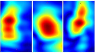
For their study, published in Scientific Reports, the team turned to high-resolution millimeter-wave imaging (HR-MMWI) — actually the same technology used for airport security screening and in some advanced 5G networks.
Healthy skin cells reflect the millimeter-wave rays differently than cancerous cells do, allowing the researchers to locate the malignant tissue. They took readings from multiple different antennas on the device and used algorithms to create a single, highly detailed image of the mole or blemish. While the waves penetrate only 2 mm into skin, that’s far enough to also create 3D models of lesions.
Healthy skin cells reflect the millimeter-wave rays differently than cancerous cells do, allowing the researchers to locate the malignant tissue.
To test the device, Stevens’ Amir Mirbeik and colleagues took a tabletop version to a clinic and used it on 71 patients during actual visits. They found that the scanner could identify cancerous tissues with 98% specificity and 97% sensitivity, numbers on par with currently used diagnostic tools.
Because the results are delivered in seconds, it could eventually help dermatologists come to more accurate diagnoses faster.
A better skin biopsy: With further development of their algorithms, the device could produce ever-better maps of lesion boundaries, allowing doctors to perform a skin biopsy that is more precise and less invasive, similar to advanced MRI imaging techniques being used in the UK for prostate cancer biopsies (which sounds way worse than my quick cut).
“We aren’t trying to get rid of biopsies. But we do want to give doctors additional tools and help them to make better decisions.”
Negar Tavassolian
The team’s next step is to shrink the technology, allowing it to be handheld and made for around $100 a pop, and hopefully ready to be used clinically within two years.
“The path forward is clear, and we know what we need to do,” Tavassolian said. “After this proof of concept, we need to miniaturize our technology, bring the price down, and bring it to the market.”
We’d love to hear from you! If you have a comment about this article or if you have a tip for a future Freethink story, please email us at tips@freethink.com.


