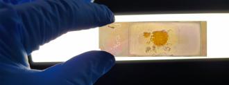
On a new smart microscope slide, breast cancer cells appear brightly colored — and that could make it easier for doctors to correctly diagnose patients when their disease is in its earliest stages, and treatments are most effective.
“Comparing images from our slides to conventional staining is like watching color television when all you’ve seen before is black and white,” co-inventor Brian Abbey from Australia’s La Trobe University said in a news release.
Spotting breast cancer cells: The sooner a patient begins treatment, the better their chance of a positive outcome. However, diagnosing breast cancer early isn’t easy — doctors must spot the relatively few cancerous cells among the many healthy ones in a tissue biopsy.
“Current approaches to tissue imaging often rely on staining or labelling cells in order to render them visible under the microscope,” Abbey said.
“Even with staining or labelling, it can be challenging for pathologists to detect cancer cells, with the risk that some samples are misdiagnosed, particularly during the very early stages of disease,” he continued.

The alternative: There is another way to detect breast cancer cells in tissue biopsies.
Due to their composition, shape, and other factors, cancer cells respond to light differently than healthy cells. This means they can be identified without stains or labels — but only if doctors use specialized microscopes and complex techniques.
Now, the La Trobe team has modified standard microscope slides so that they can reveal these visible differences in breast cancer cells without any special processing.
“[It’s] like watching color television when all you’ve seen before is black and white.”
Brian Abbey
Smart microscope slides: The NanoMslide gets this remarkable ability from a coating that manipulates how the breast cancer cells interact with light — this enhances their contrast when viewed under a standard light microscope.
Using the smart slides, the researchers could easily distinguish between breast cancer cells and healthy cells in samples of mouse and human tissue.
“When I first looked at a tissue under the microscope on the NanoMslide, I was incredibly excited,” co-lead researcher Belinda Parker said. “For the first time, I saw cancer cells just popping up at me. They were a different color from the surrounding tissue.”

Looking ahead: A new piece of equipment is also allowing the researchers to scale up production of their smart microscope slides, manufacturing batches of thousands instead of dozens.
They now plan to see if they can use those slides to detect other cancers and from other kinds of samples.
“Based on our preliminary findings with the NanoMslide, we think this platform could be really useful in early breast cancer diagnosis, but also in other cancers where we’re really just trying to pick up a few cancer cells in a complex tissue or a blood sample,” Parker said.
We’d love to hear from you! If you have a comment about this article or if you have a tip for a future Freethink story, please email us at tips@freethink.com.